By A Mystery Man Writer

Download scientific diagram | a-i Optical microscopy (first row) and FEG-ESEM (second and third rows) images of the Afghan (a, d, g), Siberian (b, e, h), and Chilean (c, f, i) lapis lazuli stones and their derived pigments (third row) from publication: Characterization of lapis lazuli and corresponding purified pigments for a provenance study of ultramarine pigments used in works of art | In this paper, we propose an analytical methodology for attributing provenance to natural lapis lazuli pigments employed in works of art, and for distinguishing whether they are of natural or synthetic origin. A multitechnique characterization of lazurite and accessory phases | Pigmentation, Paintings and Art | ResearchGate, the professional network for scientists.

Photoreceptor phagocytosis is mediated by phosphoinositide signaling - Mustafi - 2013 - The FASEB Journal - Wiley Online Library

Scanning Electron Microscopy and clay geomaterials: From sample preparation to fabric orientation quantification - ScienceDirect
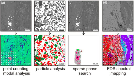
Applied Sciences, Free Full-Text
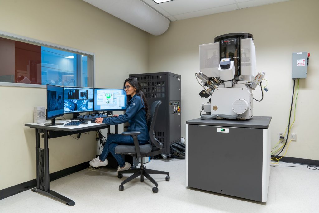
Instruments - Canadian Centre for Electron Microscopy

Applied Sciences, Free Full-Text

Scanning Electron Microscopy and clay geomaterials: From sample preparation to fabric orientation quantification - ScienceDirect

Electron-Based Imaging Techniques - ScienceDirect

PDF) Scanning electron microscopy and x-ray microanalysis-Goldstein,Newbury.pdf
a-i Optical microscopy (first row) and FEG-ESEM (second and third rows)