
A) Preoperative intraoral periapical (IOPA) radiograph of 36. B) Post operative (IOPA) radiograph of 36. C) 1 month follow up IOPA radiograph of 36. D) 6 months follow up IOPA radiograph of 36. E) 1 year follow up IOPA radiograph of 36. - IP Indian J Conserv Endod - clinical and preclinical conservative /restorative de

Radiograph sem

Incidence of periapical lesions and clinical symptoms after pulpectomy—A clinical and radiographic evaluation of 1- versus 2-session treatment - ScienceDirect

A Preoperative intraoral peri-apical (IOPA) radiograph of lower right

A) Preoperative intraoral periapical (IOPA) radiograph of 36. B) Post

a) Preoperative IOPA radiograph of tooth #36. (b) Intraoral image

Postoperative IOPA of 36 and 46.
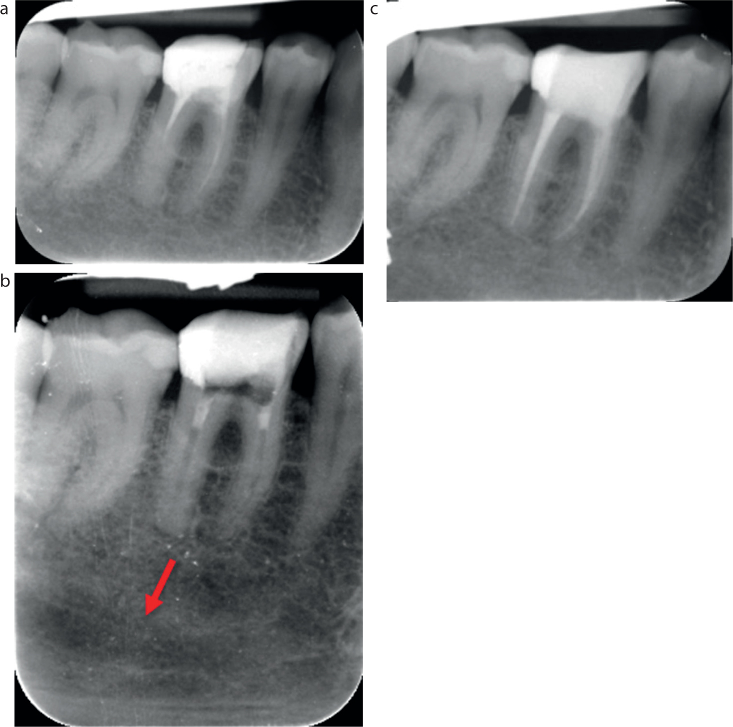
Dental Update - Assessing inferior dental nerve injury risk in

Association between concentration of active MMP‐9 in pulpal blood and pulpotomy outcome in permanent mature teeth with irreversible pulpitis – a preliminary study - Sharma - 2021 - International Endodontic Journal - Wiley Online Library
A) Preoperative intraoral periapical (IOPA) radiograph of 36. B) Post